Confluency assay

The cell responsible for the matrix deposition and contraction in all fibrotic diseases is the myofibroblast and surgically excised specimens from patients with Dupuytren's disease provide an abundant supply of material to develop assays that can be applied to other fibrotic conditions where primary early disease stage human tissues are less readily available. Myofibroblasts interact with the local extracellular cellular matrix (ECM) and sense tissue forces through transmembrane ECM receptors such as integrins. Upon adhesion to the ECM, integrins recruit intracellular proteins to form a focal adhesion (FA), which links integrins to the actin cytoskeleton and initiates force-dependent signaling. The ability to spread and interaction with the ECM is critical to myofibroblast function.
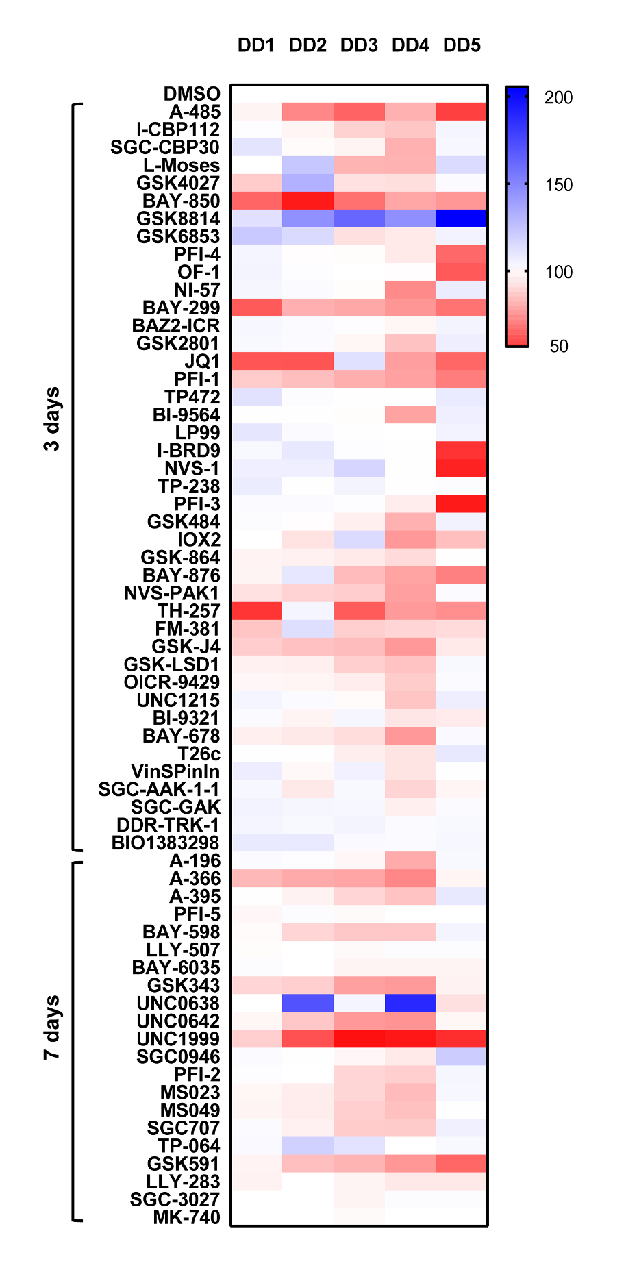
We set up the confluency assay to rapidly screen the ability of cells to spread and interact with the ECM. Using surgically excised tissue from Dupuytren’s patients we derived early passage myofibroblast2 (p2) which are the basis for all our assays. Cells are allowed spread and adhere then treated with probes for 3 days in the present of Calcein (cell-permanent fluorescent dye). The effect of the probe set on confluency is detected and quantified using Celigo image cytometer.
The effects of the probe set on cell confluency were most analogous to the cell proliferation data. A485 (acetyltransferase), BAY-850 (ATAD2), BAY-299 (BRD1), the BRD4 probes JQ1 and PFI-1, the methyltransferase inhibitors A-366 (G9a/GLP), GSK343 (EXH2), UNC0642 (G9a/GLP) and UNC1999 (EXH2) all showed similar effects on confluency. However with the exception of the PRMT5 probe (GSK591) none of the probes targeting the PRMT family showed any major effects on confluency. TH-257 the LIMK inhibitor did show marked effects on confluency without any effect on viability and displayed only limited effects on proliferation. Given the role of LIM kinases in microtubule assembly this would confirm this assay is able to detect probes which can modify actin cytoskeleton/cell spreading phenotype in the myofibroblasts.
- Surgically excised tissue from Dupuytren’s patients was treated with collagenase to derive single cell suspensions; cells were passaged (p2) and are termed myofibroblasts.
- Cells were seeded in 96 wells at 2 x10³/well and left to adhere overnight. The following day probes were added at 1µM in triplicate, using DMSO as the vehicle control. Probes were incubated with the cells for 3 days or in the case of methyltransferase (MT) probes 7 days (cells were treated with MT probes again at day 3).
- 1 hr prior to the end of the assay, calcein (250ng/ml, final concentration) was added to microplate wells.
- Quantitative cell counting was performed using the Celigo image cytometer. Data is expressed as % compared to DMSO control (100%), DD1-5 Dupuytrens’s disease patients.
Cell Confluency